Nuclear medicine uses nuclear imaging to help diagnose and monitor various bone diseases. The process involves small amounts of radioactive substances, called radiotracers, and imaging equipment to provide detailed pictures of bones inside the body. These tests help doctors identify causes of bone pain and track bone changes that may not be visible on standard X-rays.
Understanding Nuclear Medicine
Nuclear medicine is an advanced diagnostic approach. It uses radiotracers, which are tiny amounts of radioactive materials. These may be injected into a vein, taken orally, or used by other methods, depending on the procedure. The radiotracers travel through the bloodstream and accumulate in certain organs or tissues. Special cameras detect the radioactivity from these tracers and produce images for review.
This imaging technique focuses on bone metabolism and changes in the structure of bones. As the radiotracer is absorbed more by actively changing or abnormal bone tissue, the resulting images can help detect bone conditions. Nuclear medicine differs from regular X-rays, which primarily show bone structure and density.
Diagnostic Procedures Included
A bone scan is a common type of nuclear medicine exam used to evaluate the entire skeletal system. The scan can help find the source of unexplained bone pain, diagnose fractures, detect arthritis, find bone infections, and monitor cancer in bones. A bone scan may also detect cancer that has spread from another area.
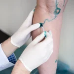
During a bone scan, a doctor or technologist injects a radiotracer into a vein in your hand or arm. The tracer takes between two and four hours to circulate and be absorbed by bones. Sometimes, images are taken soon after the injection; main imaging is usually done a few hours later. Patients may be asked to drink several glasses of water to help clear unabsorbed tracer from the body. You will be asked to empty your bladder before scanning to improve image quality.
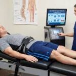
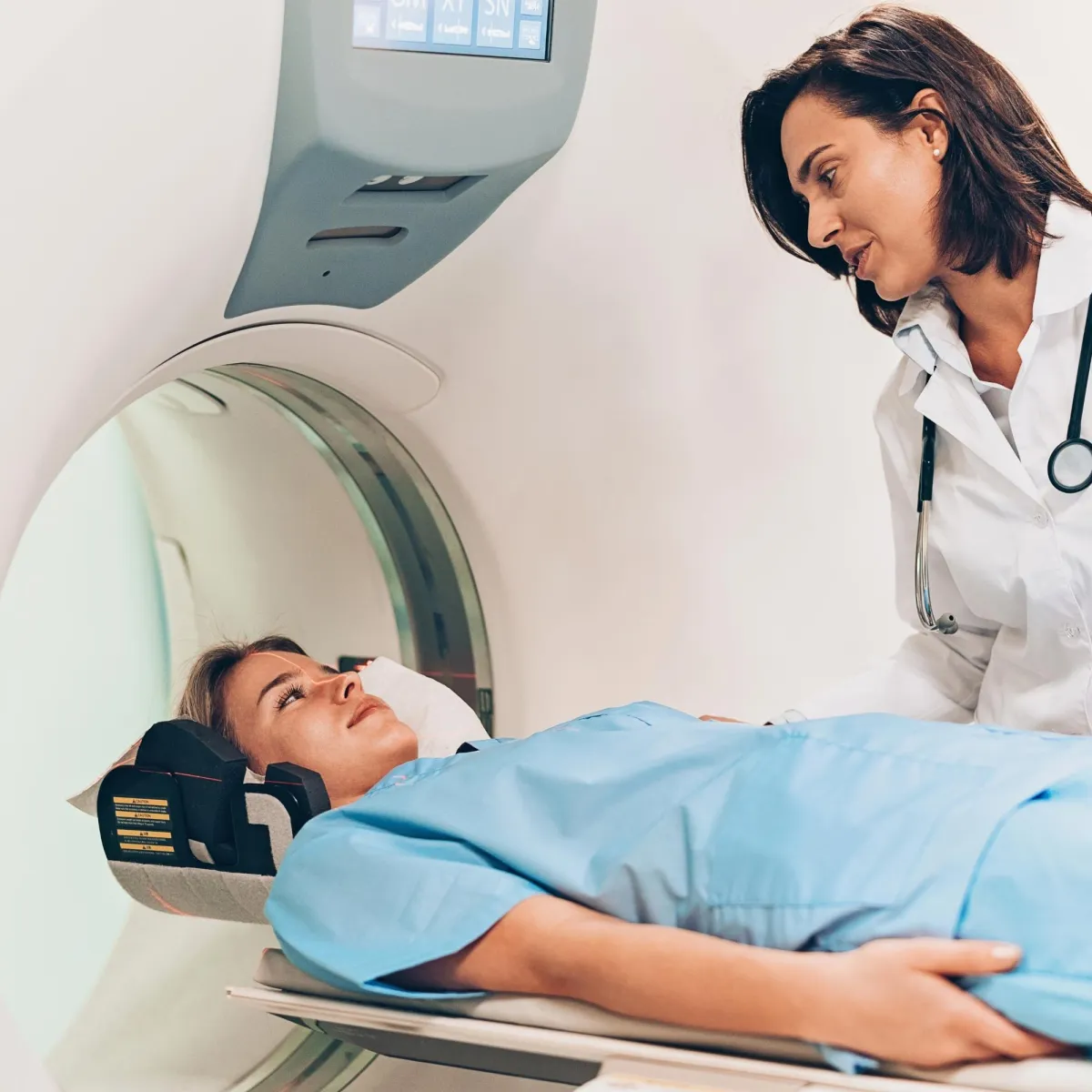
For the scan, the patient lies still on a table while a camera passes over the body to detect tracer emissions. The procedure is painless and takes up to an hour. In some cases, the doctor may order a three-phase bone scan or additional imaging, such as single-photon emission computerized tomography (SPECT), for more detailed evaluation of difficult-to-see areas.
Post-Procedure Expectations
Most people can return to normal activities right after a bone scan. The procedure generally has no side effects and involves minimal radiation exposure—less than a typical CT scan. The radioactivity from the tracer leaves the body mainly through urine; patients are usually encouraged to drink plenty of water for a day or two following the exam. The tracer is usually gone within two days after the scan.
Doctors provide instructions for the period after the scan. Women who are pregnant or nursing should tell their doctor before undergoing a nuclear medicine procedure; these scans are not usually performed during pregnancy or breastfeeding due to the potential risk of radiation to the baby.
A radiologist reviews the bone scan images and sends a report to the referring doctor. The results identify areas of unusual bone metabolism. These appear as “hot spots” (where more tracer is absorbed) or “cold spots” (where less tracer is present). Further testing might be needed if abnormal areas are found, since a bone scan is sensitive to changes but may not determine the exact cause.
Find a Clinic
Nuclear medicine, especially bone scans, provides doctors with valuable details to assess bone disease and injury. The process uses radiotracers and specialized imaging to evaluate changes in bone metabolism. Bone scans are used for various clinical reasons, including diagnosing fractures, infections, arthritis, or cancer affecting the bones. These procedures are safe and informative, offering guidance for further care when bone disease is suspected. Find a clinic near you to undergo testing.